A new capsule endoscopy system (OMOM), equipped with artificial intelligence, supports the diagnosis and treatment of patients at the Gastroenterology Clinic of the University Hospital “Tsarina Ioanna – ISUL“. Artificial intelligence is trained to detect even the smallest changes in the mucous membrane of the gastrointestinal tract and mark them on the corresponding photos. The program also has the ability to determine the characteristics of the marked findings – for example – whether they are ulcers, polyps or something else. In this way, with the help of new technologies, the frequency of missed diseases is reduced and the time for reading the results is significantly reduced. FACTI spoke to Dr. Viktor Dimitrov, one of the doctors who performs the high-tech examination at ISUL.
- Dr. Dimitrov, what is “smart“ capsule endoscopy and how does it differ from traditional gastroscopy or colonoscopy?
- The epithet “smart“, often used in the context of capsule endoscopy, in this case embodies the presence of software with the help of which endoscopically visible diseases are caught in the anatomical areas mirrored by the capsule. This software essentially represents artificial intelligence, which is trained with numerous photo materials from real and proven diseases to recognize the characteristics of most mucosal disease processes. It has the ability to detect and characterize the relevant finding. Its extremely useful quality is to filter out repetitive and insignificant frames from the recording, thus significantly reducing the reading time. These AI skills have been validated with clinical studies verifying high levels of specificity, sensitivity and accuracy.
It differs from conventional endoscopic examinations in its potential to adequately examine the length of the small intestine, which remains inaccessible to gastroscopy and colonoscopy.
It is a useful tool in patients with an unclear source of bleeding, whether manifest (clear blood or melena in stool) or occult (unexplained iron deficiency anemia), in whom gastro- and colonoscopy have already been performed - without finding an explanation for the hemorrhage (bleeding). It should be noted that capsule endoscopy does not replace gastro- and colonoscopy for examination of the stomach and colon. The inability to take a biopsy from a disease documented by the capsule in itself indicates the performance of conventional endoscopic examinations. Colonoscopy is a safe intervention and the gold standard for screening for colorectal cancer, and capsule endoscopy is recommended only for certain contraindications to colonoscopy. I emphasize the last sentence because patients often desire capsule endoscopy due to stigma or fear of colonoscopy, which are in most cases unjustified.
- The study was first introduced in Bulgaria at ISUL almost a year ago. How many patients have you examined with this innovative approach so far?
- We have conducted nearly 50 studies so far, and it should be noted that patients are largely selected strictly according to serious indications, in line with modern international recommendations. It is rare to perform capsule endoscopy on a patient who has not undergone conventional endoscopic examinations without having contraindications for them.
- Is this examination method suitable for every patient?
- Of course not. After a preliminary consultation, it is assessed whether the patient has a real indication for performing capsule endoscopy, and the available contraindications are necessarily taken into account. Everything is discussed with the patient and the benefits, risks and meaning are explained in each individual case.
- For which patients is capsule endoscopy contraindicated?
- According to current international data, the main real contraindication for capsule endoscopy is the risk of capsule retention — that is, in patients with known or strongly suspected mechanical obstruction, stricture, or significant stenosis of the small intestine, as well as in some fistulas, when there is a real risk that the capsule will not pass. In patients with symptoms of subileus/ileus (obstruction) or known stenosis (narrowing), imaging of the small intestine (computed tomography or magnetic resonance enterography) should first be performed.
Swallowing disorders are not an absolute contraindication, but they are an important relative one.
In patients with dysphagia (difficulty swallowing), neurological disease, patients with impaired swallowing reflex or patients with previous aspiration, there is a risk of the capsule getting into the respiratory tract. This complication is rare, but real and requires careful selection. In such patients, the capsule can be placed endoscopically in the duodenum (duodenum) instead of being swallowed.
Pregnancy itself is considered a relative contraindication, not because there is a proven high harm, but because safety data are limited. Modern reviews and practical recommendations assume that capsule endoscopy should be performed in pregnant women only when there is a strong clinical need, when the result will change behavior and the examination cannot necessarily be postponed until after delivery.
- What are the diseases that are “looked for“ with the help of capsule endoscopy in general and in particular the one that uses artificial intelligence?
- The most common indication for which capsule endoscopy is used, as already pointed out, is manifest or occult bleeding of unclear origin, not specified by conventional endoscopic examinations already performed. In this context, angiodysplasias, small ulcerative lesions or rare vascular anomalies are mainly sought, but also neoplastic processes (neoplastic formations that can cause cancer). In this group, capsule endoscopy has the highest diagnostic potential and is practically the method of choice.
The second large group of patients are those with suspected or proven Crohn's disease,
especially when it comes to early disease or proximal involvement, in conventionally endoscopically inaccessible parts of the small intestine. Capsule endoscopy has been shown to be suitable for detecting mucosal lesions such as aphthous ulcers, erosions and the typical skipping lesion, making it a more sensitive method than imaging studies for early inflammatory changes. It is also used in disease monitoring and assessment of mucosal healing, although in patients with suspected stenosis the risk of capsule retention should always be assessed.
Capsule endoscopy also has an essential role in the diagnosis of small bowel tumors, which are rare but often present with nonspecific symptoms such as anemia or weight loss. It can visualize adenocarcinomas, neuroendocrine tumors, GIST, lymphomas and metastases, and is often the first method to point to diagnosis. In addition, it is used in patients with hereditary polyposis syndromes such as Peutz-Jeghers or familial adenomatous polyposis to assess the number and distribution of polyps in the small intestine, as well as in complicated or refractory celiac disease, where it can detect characteristic morphological changes or complications such as ulcerative jejunitis and enteropathy-associated T-cell lymphoma.
With the advent of artificial intelligence, the spectrum of diseases that are sought does not change significantly, but rather the way in which they are detected and interpreted.
AI (artificial intelligence)-assisted capsule endoscopy significantly improves lesion detection, with the best validated algorithms for recognizing blood and vascular abnormalities, where the sensitivity of the study is over 95-99%.
Also, AI demonstrates very good results in detecting ulcers and erosions, making it particularly useful in patients with suspected Crohn's disease or NSAID-induced enteropathy. In polyps and tumors, the results are also promising, but still more variable, as these lesions are morphologically more heterogeneous.
One of the most important practical benefits of AI in capsule endoscopy, as previously highlighted, is its ability to filter a large volume of normal images and mark “suspicious“ frames, which significantly reduces the reading time and the risk of missing small but clinically significant lesions.
However, at present, artificial intelligence functions as a highly effective detection tool, but cannot replace clinical interpretation, especially in terms of assessing the significance of the findings, differential diagnosis and their integration into the overall clinical context. In this sense, the most modern and optimal approach is a combined – using AI to increase diagnostic sensitivity and efficiency, but with a final expert assessment by a specialist physician.
It is pertinent to add that training is needed for personnel handling AI, as there are potential negatives and shortcomings of AI, which, if not recognized and actively avoided, lead to compromising the effectiveness of the method. In the Gastroenterology Clinic of UMBAL “Tsarina Ioanna – ISUL“, where artificial intelligence has been actively used for more than a year, the interpretation of the results is carried out not only by me, but also by Dr. Hristo Valkov and Dr. Mila Kovacheva, all three of whom have undergone special training for this purpose.
- Is the examination covered by the NHIF?
- Yes, the examination is covered by the NHIF, after a specialist has assessed the presence of an adequate indication and accordingly requires a short stay in a medical facility.
- We have talked a lot about colon cancer, which remains one of the main causes of fatal outcomes for patients in Bulgaria. In our country, the disease occurs many times more often than in Western European countries. Why is this so?
- Based on the most current and relatively reliable international data, it cannot be convincingly stated that colorectal cancer is more common in Bulgaria than in Western Europe. It is more accurate to say that Bulgaria has a distinct and probably underestimated burden of the disease, but not a clearly proven higher incidence compared to Western Europe. The reason is that the latest OECD/European Commission profile for Bulgaria indicates that the registered total cancer incidence in the country is low compared to the EU average, and this is largely due to a currently non-functional registry that is unable to report the real incidence of colorectal cancer, accompanied by the lack of a national colorectal cancer screening program. A planned pilot program should be launched on the issue, but the facts will be clear when they are actually implemented.
- Many people are worried about colonoscopy, which is considered the gold standard for the diagnosis and prevention of colon cancer, because the examination is invasive and not particularly pleasant. Is modern medicine working towards inventing an alternative that does not scare patients so much?
- This is not a problem of modern medicine, but a problem of the general health culture in Bulgaria. There are indeed many patients who, for reasons unknown to us, present themselves with an extremely negative attitude towards colonoscopy, and the arguments are rarely based on real risks and contraindications. It should be known that colonoscopy is, above all, the best method for screening and prevention of colorectal cancer and should not, in the absence of real contraindications, tolerate an alternative. It is a largely safe intervention, and when anesthesia is applied, it is also painless. In the event of a contraindication to anesthesia or at the patient's request, colonoscopy without anesthesia, with proper technique, should not cause significant pain, unless, of course, there are accompanying intestinal diseases or previous surgical interventions that would complicate the examination and, accordingly, cause more pronounced pain.
Colonoscopy without anesthesia has its positive sides, but this is a topic for another discussion. I would like to reassure patients that colonoscopy is in a very high percentage without significant discomfort, especially under anesthesia, where discomfort is minimal or completely absent. I would like to note that there are quite a few patients who, after having the examination without anesthesia, no longer want anesthesia for subsequent colonoscopy, as they are convinced that there is no reason to fear - here, the leading factors are undoubtedly the anatomical features, the underlying diseases, the colonoscopy technique and who performs it. We don't need a less intimidating method, but more accessible information for the patient, presented in a clear and understandable form.
- Are there specific symptoms that you doctors use to distinguish whether it is a problem in the small or large intestine? What should patients watch for?
- In medicine, there is rarely a symptom that can definitely indicate whether the problem is in the small or large intestine. However, there are certain guidelines that we follow, always considering them in the context of the overall clinical picture. In general, when it comes to problems in the small intestine, we more often see symptoms related to impaired absorption of food — for example, chronic diarrhea, bloating, gas, weight loss or vitamin and iron deficiency. The pain is usually more diffuse, around the navel, and is not so clearly localized.
On the other hand, diseases of the large intestine more often manifest themselves with changes in bowel movements — alternating constipation and diarrhea, change in stool form, presence of mucus or blood, as well as more localized pain in the lower abdomen.
But it is important to emphasize that these are not strict rules. For example, blood in the stool can also come from higher levels of the digestive tract, and diarrhea can be positive in diseases of both the small and large intestines. Therefore, a diagnosis is not made based on symptoms alone, but tests such as endoscopy, imaging diagnostics or capsule endoscopy are used to specify the source of the problem.
The most important thing for patients is to monitor for so-called alarming symptoms — such as unexplained weight loss, blood in the stool, prolonged diarrhea or constipation, anemia — and to seek medical help in a timely manner.
-----------------------------------
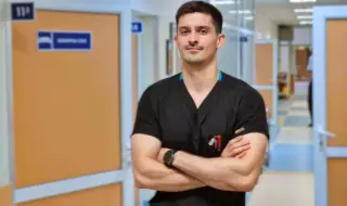
Dr. Viktor Dimitrov graduated from MU-Sofia in 2022, after which he became part of the team of the Gastroenterology Clinic at UMBAL “Tsarina Ioanna-ISUL". Since 2023, he has been an assistant professor at the Department of Gastroenterology at MU-Sofia. He participates in national and international conferences on gastroenterology and hepatology. He has completed training and courses abroad in the field of interventional gastroenterology. He is actively involved in endoscopic manipulations of the hepatobiliary system and emergency interventional endoscopy in cases of bleeding from the gastrointestinal tract.